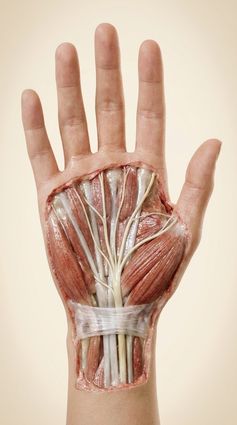
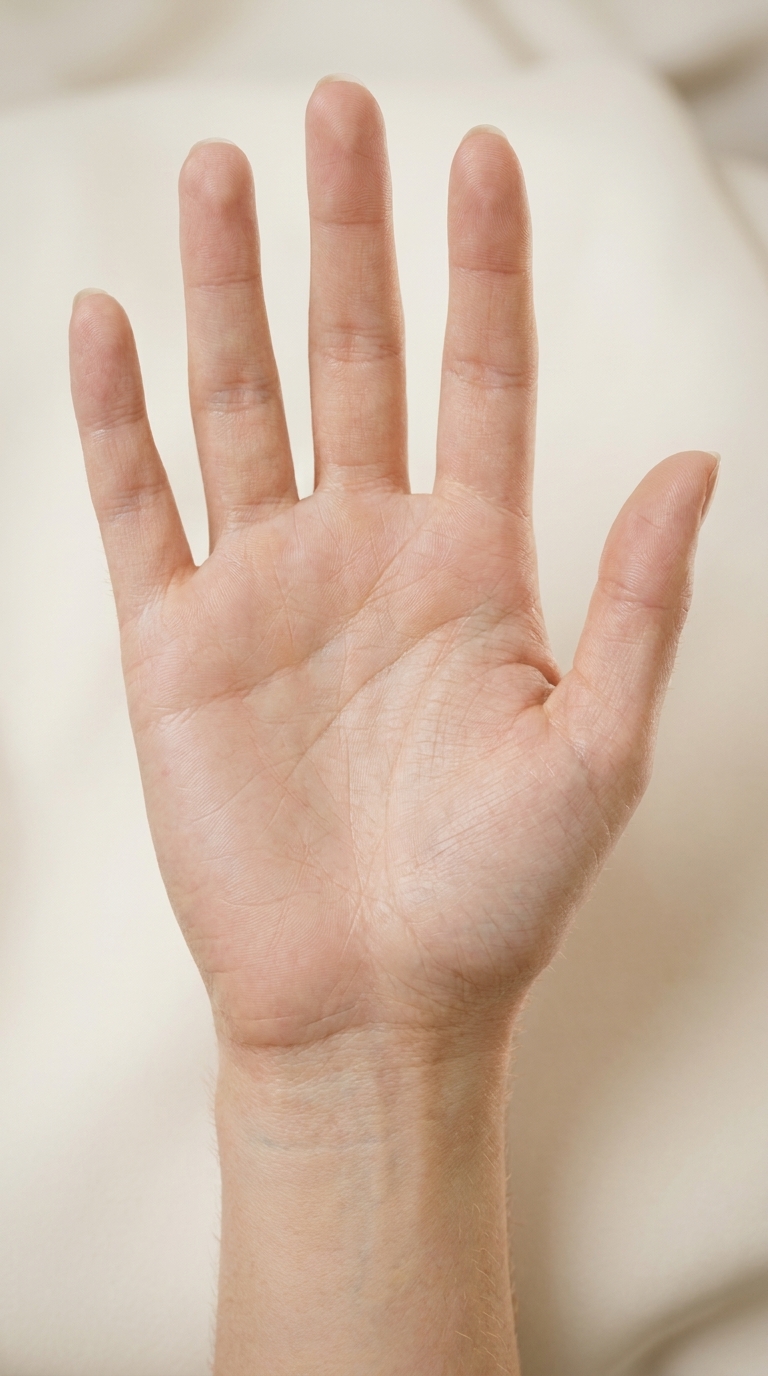
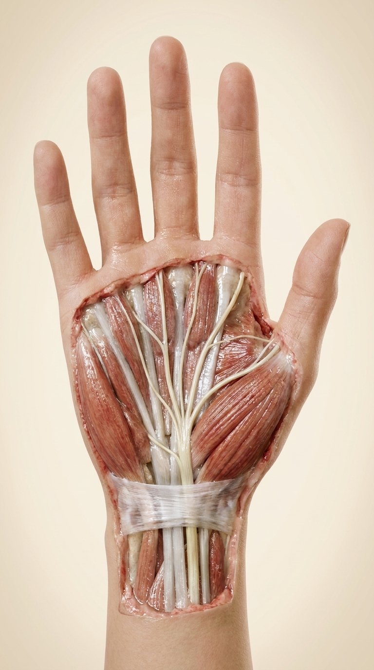
Hyper-photorealistic anatomical dissection of the same right palm and wrist, palm up, same framing and position as reference 1, on a soft cream background. Skin and palmar fascia removed at the wrist and palm, real wet cadaver-like dissection photographed in a medical atlas style: glistening tendons with visible fibres, flexor retinaculum (transverse carpal ligament) as a white fibrous band, the median nerve as a pale yellow-white cord with fine longitudinal fascicles passing under the retinaculum through the carpal tunnel, then branching to thumb, index, middle and the radial half of the ring finger, matching reference 2. Thenar muscles reddish with fibre texture. Skin still intact on the fingers, cut edge realistic. Macro photography, soft studio light. Five fingers, anatomically correct. No text, no labels.